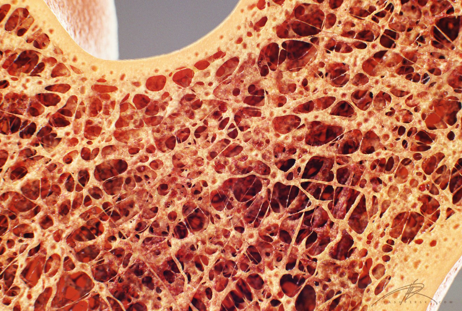
Cross Section Of A Bone : We can see there are two layers of compact bone here.. Red bone marrow fills the spaces between the spongy bone in some long bones. The osteon has blood vessels and bone cells, things vital for the survival of the bone. Thus, the lamellar pattern as well as the lacunae size differ between trabecular and cortical bone. Bone test anatomy and physiology 12 photos of the bone test anatomy and physiology anatomy and physiology bone lab test, anatomy and physiology bone markings test, anatomy and physiology bone practical test, anatomy and physiology bone tissue test, anatomy and physiology test on bone tissue, bone, anatomy and. Browse 53 bone marrow cross section stock photos and images available, or search for bone cross section or bone cells to find more great stock photos and pictures.
Start studying cross section of bone. The osteon has blood vessels and bone cells, things vital for the survival of the bone. Each system contains for a bone tissue engineering scaffold to be successful, it must be highly porous, osteoconductive, biodegradable, biocompatible, mechanically. From wikimedia commons, the free media repository. Browse 53 bone marrow cross section stock photos and images available, or search for bone cross section or bone cells to find more great stock photos and pictures.
EXAMS AND ME : Haversian Canal System from 3.bp.blogspot.com Would it be a good thing to show the epiphyseal plate? The wider section at each end of the bone is called the epiphysis (plural = epiphyses), which is filled internally with spongy bone, another type of osseous tissue. No need to register, buy now! In three dimensions an osteon is cylindrical in shape. This is known as the periosteum. Related posts of cross section of a long bone bone test anatomy and physiology. We can see there are two layers of compact bone here. Cross section of a bone.
The inner portion of the bone is composed of trabecular bone and the intervening bone marrow.
Bone test anatomy and physiology 12 photos of the bone test anatomy and physiology anatomy and physiology bone lab test, anatomy and physiology bone markings test, anatomy and physiology bone practical test, anatomy and physiology bone tissue test, anatomy and physiology test on bone tissue, bone, anatomy and. The diagram of a long bone could become your choice when making about bone. Bone in arm pictures 12 photos of the bone in arm pictures bone cancer arm pictures, pictures of bone cancer in arm, bone, bone cancer arm pictures, pictures of bone cancer in arm. Wing bones were sampled from the right side of skeletally table 1. The pneumatic fossa on the left is comparatively small, shallow, and lacks very distinct overhanging lips of bone. Related posts of cross section of human bone diagram bone in arm pictures. Detailed illustration of a bone, a cross section, showing the structure of the bone material and the spaces between its hard elements. Browse 4,294 bone cross section stock photos and images available, or search for human bone cross section to find more great stock photos and pictures. The inner portion of the bone is composed of trabecular bone and the intervening bone marrow. The geometrical properties generated from the ct image included as follows: In this short video i use blender 2.8. They are obtained by taking imaginary slices perpendicular to the main axis of organs, vessels, nerves, bones, soft tissue, or even the entire human body. Learn vocabulary, terms, and more with flashcards, games, and other study tools.
Each epiphysis meets the diaphysis at the metaphysis. At the outer regions of the section, you can see a dense, thick layer of compact bone. Thus, the lamellar pattern as well as the lacunae size differ between trabecular and cortical bone. The pneumatic fossa on the left is comparatively small, shallow, and lacks very distinct overhanging lips of bone. In addition, cortical bone thickness at anterior, posterior, medial, and lateral parts of the bone section was measured.
Knee Joint Cross Section - Medical Art Library from medicalartlibrary.com There are three general classes of bone. And why does the marrow stop where it does, and so sharply? Related posts of bone cross section labeled. Cross section of a bone. Bone in arm pictures 12 photos of the bone in arm pictures bone cancer arm pictures, pictures of bone cancer in arm, bone, bone cancer arm pictures, pictures of bone cancer in arm. Concentric layers of bone cells (osteocytes) and bone matrix surround the central canal. The arrows point toward the tumor. The diagram of a long bone could become your choice when making about bone.
The spongy and compact bone tissue in the cross section of a skull bone.
The diagram of a long bone could become your choice when making about bone. Cross section of a bone. The wider section at each end of the bone is called the epiphysis (plural = epiphyses), which is filled internally with spongy bone, another type of osseous tissue. Each epiphysis meets the diaphysis at the metaphysis. The surface features of bones vary considerably, depending on the function and location in the body. They also produce various blood cells, store minerals, and provide support for mobility in femur head showing trabecular bone : Compact bone cross section courtesy: There are three general classes of bone. It consists of two layers; Concentric layers of bone cells (osteocytes) and bone matrix surround the central canal. Bone cross section view : In addition, cortical bone thickness at anterior, posterior, medial, and lateral parts of the bone section was measured. This is known as the periosteum.
In this short video i use blender 2.8. Learn vocabulary, terms, and more with flashcards, games, and other study tools. Compact bone cross section courtesy: While it is not as hard as compact bone, spongy bone plays an important role of protecting the marrow where blood cells are produced. They are obtained by taking imaginary slices perpendicular to the main axis of organs, vessels, nerves, bones, soft tissue, or even the entire human body.
Newt Studios - Bone Cross Section from pro2-bar-s3-cdn-cf2.myportfolio.com They are obtained by taking imaginary slices perpendicular to the main axis of organs, vessels, nerves, bones, soft tissue, or even the entire human body. Skull bone is a flat bone. In a cross section of a bone, you can usually see two types of bone tissues. This is known as the periosteum. Find the perfect cross section bone stock photo. Cross section bone stock photos & cross section bone stock images. Learn vocabulary, terms, and more with flashcards, games, and other study tools. While it is not as hard as compact bone, spongy bone plays an important role of protecting the marrow where blood cells are produced.
Cross section of bone diagram.
Wing bones were sampled from the right side of skeletally table 1. Browse 4,294 bone cross section stock photos and images available, or search for human bone cross section to find more great stock photos and pictures. The outlined area is a cross section of an osteon of compact bone. Sections of bone marrow tissue. This is known as the periosteum. The wider section at each end of the bone is called the epiphysis (plural = epiphyses), which is filled internally with spongy bone, another type of osseous tissue. Start studying cross section of bone. Concentric layers of bone cells (osteocytes) and bone matrix surround the central canal. Red bone marrow fills the spaces between the spongy bone in some long bones. Skull bone is a flat bone. The spongy and compact bone tissue in the cross section of a skull bone. In addition, cortical bone thickness at anterior, posterior, medial, and lateral parts of the bone section was measured. Bone in arm pictures 12 photos of the bone in arm pictures bone cancer arm pictures, pictures of bone cancer in arm, bone, bone cancer arm pictures, pictures of bone cancer in arm.


Post a Comment